

重组抗小鼠VEGF单抗(G6-31) | Syd Labs PA007515.m2a
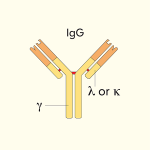
体内实验级重组抗小鼠VEGF单克隆抗体(克隆号:G6-31,小鼠IgG2a kappa,货号:PA007515.m2a)是用哺乳动物细胞生产的重组抗体,适用于体外和体内研究,纯度>95%。Syd Labs PA007515.m2a抗小鼠VEGF抗体不变区为小鼠Mouse IgG2a Kappa (mIgG2a或m2a),可与重组小鼠IgG2a同型对照抗体配套使用。样品制备条件和最佳样品稀释度应由研究人员通过实验确定。

产品参数
| 货号 | PA007515.m2a |
|---|---|
| 产品名称 | 重组抗小鼠VEGF单抗(G6-31) | Syd Labs PA007515.m2a |
| 英文名 | In Vivo Grade Recombinant Anti-mouse VEGF Monoclonal Antibody (Clone: G6-31), Mouse IgG2a Kappa |
| 供货商名称 | Syd Labs, Inc. |
| 品牌名 | Syd Labs |
| 别称 | 血管内皮生长因子、VEGF |
| 概述 | Syd Labs提供重组小鼠IgG2a同型对照抗体。样品制备条件和最佳样品稀释应由研究人员通过实验确定。 |
| 克隆号 | G6-31 |
| 同种型 | 小鼠 IgG2a kappa |
| 特异性 | 体内实验级重组小鼠单克隆抗体(克隆:G6-31)特异性结合小鼠VEGF |
| 应用 | ELISA、中和、功能测定,如生物分析PK和ADA测定,以及用于研究小鼠VEGF蛋白影响的生物途径的测定。 |
| 抗体形式 | 0.2 μM过滤溶液,1x PBS |
| 内毒素 | 根据 LAL 方法,≤1 EU每1mg 蛋白质 |
| 纯度 | >95%(在还原条件下通过SDS-PAGE测定) |
| 运输 | 体内实验级重组抗小鼠VEGF单克隆抗体,小鼠IgG2a kappa(克隆号G6-31)用冰袋运输。收到后,请立即将其存放在下面建议的温度下。 |
| 稳定性与存储 | 使用手动除霜冰箱并避免重复冻融循环。 自收到之日起 1 个月,保存在2 至 8°C。 自收到之日起12个月,保存在-20 至 -70°C。 |
| 注意事项 | PA007515.m2a Syd Labs:体内实验级重组抗小鼠VEGF小鼠IgG2a kappa单克隆抗体是用哺乳动物细胞生产的重组抗体,可与重组小鼠IgG2a同型对照抗体配套使用。样品制备条件和最佳样品稀释应由研究人员通过实验确定。 |
| 产品咨询 | Syd Labs在国内只通过代理商销售其产品,不做直销。终端用户咨询价格请联系Syd Labs中国代理商。 关于Syd Labs产品如果有任何技术或其它问题,欢迎随时联系Syd Labs国内市场推广合作伙伴:武汉多找找科技有限公司,企业微信:duozhaozhao2024 联系电话:18162581039(龙经理) |
| 应用详情 | ELISA、中和、功能测定,如生物分析PK和ADA测定,以及用于研究小鼠VEGF蛋白影响的生物途径的测定。 |
文献
PA007515.m2a:体内实验级重组抗小鼠VEGF单克隆抗体(克隆号G6-31,小鼠IgG2a kappa)
体内实验级重组抗小鼠VEGF单克隆抗体(克隆号G6-31,小鼠IgG2a Kappa)是针对小鼠血管内皮生长因子(VEGF/VEGF-A)的特异性阻断试剂。VEGF作为促血管生成的核心细胞因子,在肿瘤血管生成、血管通透性调节及肿瘤微环境重塑中发挥关键作用。G6-31克隆号抗体能高亲和力结合小鼠及大鼠VEGF,广泛应用于肿瘤生长抑制、眼部血管新生模型、流式细胞术(FC)及体外中和分析,是抗血管生成治疗与肿瘤免疫联合疗法研究的经典工具。
作为高品质的体内实验级重组抗小鼠VEGF单克隆抗体(克隆号G6-31),该产品采用基因工程重组表达与小鼠IgG2a Kappa骨架设计,彻底消除了传统杂交瘤生产中的批次间差异,并具有极高的一致性与特异性。其低内毒素(<1 EU/mg)与无叠氮化钠配方可有效避免非特异性毒性反应,非常适合用于长期动物体内实验(In Vivo)、血管生成机制探究及联合靶向药效评估,为高质量科研数据提供有力保障。
抗小鼠VEGF单抗(G6-31)部分参考文献:
Katsuhiro Miyajima, et al. Acta Histochem Cytochem. 2010. PMID: 20514290
“To investigate whether tumor development in E2-induced PRLoma is inhibited by anti-VEGF monoclonal antibody (G6-31), we evaluated tumor growth and observed the vascular structures. With simultaneous treatment with G6-31 for the latter three weeks of the 13-week period of E2 stimulation (E2+G6-31 group), the following inhibitory effects on the PRLoma were observed in the E2+G6-31 group as compared with the E2-only group. In the E2+G6-31 group, a tendency to reduction in pituitary weight was observed and significant differences were observed as (1) reductions in the Ki-67-positive anterior cells, (2) increases in TUNEL-positive anterior cells, and (3) repair of the microvessel count by CD34-immunohistochemistry.”
2. Sprouting Angiogenesis in Human Pituitary Adenomas.
Jie Zhou, et al. Front Oncol. 2022. PMID: 35600354
“Anti-VEGF treatment has been demonstrated to successfully treat pituitary tumors in mouse models. Korsisaari et al. used the anti-VEGF antibody G6-31 to treat pituitary tumors in Men1+/- mice (53). After 67 days, they observed that G6-31 treatment reduced the tumor volume by 72% compared to the control group and significantly extended their survival. All pituitary tumors in Men1+/- mice were later identified as prolactinomas (53). The G6-31 antibody was also used to treat Drd2(-/-) mice with hyperplastic pituitary glands by Luque et al. (54).”
Samuel A Blackman, et al. bioRxiv. 2025. PMID: 41306516
“This can occur via multiple mechanisms, including upregulation of placental growth factor 2 (PlGF-2), an alternative ligand for VEGF receptor 1 (VEGFR1) and neuropilin receptor 1 (NRP1). Activity of both growth factors is mediated by interactions with multiple receptors and extra-cellular matrix components, which complicates efforts to understand their contributions to cancer progression. To complement existing antibodies, we discovered those blocking interactions between PlGF-2 or VEGFA and their shared NRP1 receptor in the presence of heparin. Limiting angiogenesis to promote vascular normalization is one mechanism of anti-VEGF protection; here, anti-VEGFA antibodies blocking interactions with VEGFR1 and NRP1 reduced HUVEC tube formation in a physiological angiogenesis model.”
您可能还会喜欢…
-
阅读更多Quick View