
重组抗小鼠/人CD44单抗(IM7.8.1) | Syd Labs PA007622.r2b

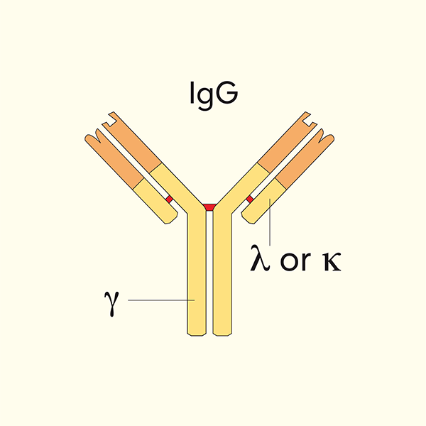
Syd Labs体内实验级重组抗小鼠/人 CD44 单克隆抗体,克隆号 IM7.8.1,恒定区为大鼠 IgG2b Kappa(Rat IgG2b Kappa),由哺乳动物细胞重组表达,跨种属识别小鼠与人 CD44 分子。CD44 又称 CD44抗原前体(CD44 antigen precursor)、CDw44、ECMR-III(细胞外基质受体III)、Epican(表皮细胞黏附分子)、Hermes抗原、Pgp-1(吞噬糖蛋白-1)、H-CAM、HUTCH-I、透明质酸受体(Hyaluronan/HA receptor),是一种 80–95 kDa I 型跨膜糖蛋白(基因名 Cd44 小鼠2号染色体、人类11号染色体11p13),胞外含 N 端 Link 结构域(透明质酸 HA 结合位点)、可变剪接区(v1–v10)、跨膜区和胞质尾,广泛表达于所有白细胞(T/B/NK/单核/中性粒细胞)、内皮细胞、肝细胞、间充质细胞、成纤维细胞及多种肿瘤细胞,作为细胞外基质成分透明质酸(HA,Hyaluronic acid)的主要细胞表面受体,参与细胞-细胞外基质黏附、白细胞滚动黏附、淋巴细胞归巢至炎症部位、造血祖细胞-骨髓基质互作、T 细胞共刺激、肿瘤转移、炎症细胞招募等过程;活化 T 细胞与记忆 T 细胞(尤其是 Tmem、Treg)表面 CD44 高表达,是流式区分 naive T(CD44low)与记忆/效应 T(CD44high)的经典标志。IM7.8.1 是 Trowbridge 等建立、识别 CD44 恒定区 N 端透明质酸结合结构域的经典大鼠单克隆抗体,可阻断 CD44-HA 相互作用,体内外广泛用于阻断 CD44 介导的细胞黏附、炎症细胞招募、自身免疫性关节炎(CIA)、造血、肿瘤侵袭转移及 CD44 信号通路研究。本品经 Protein G 亲和纯化,还原性 SDS-PAGE 及 HPLC 检测纯度>95%,LAL 法内毒素<1 EU/mg,0.2 μM 过滤于 pH 7.4、不含稳定剂或防腐剂的 1× PBS 溶液,适用于 ELISA、中和/阻断实验、功能实验(如生物分析 PK 与 ADA 检测)以及受小鼠/人 CD44 蛋白影响的生物学通路研究和体内动物实验。
产品参数
| 货号 | PA007622.r2b |
|---|---|
| 产品名称 | 重组抗小鼠/人CD44单抗(IM7.8.1) | Syd Labs PA007622.r2b |
| 英文名 | In Vivo Grade Recombinant Anti-mouse/human CD44 Monoclonal Antibody (Clone: IM7.8.1), Rat IgG2b Kappa |
| 供货商名称 | Syd Labs, Inc. |
| 品牌名 | Syd Labs |
| 别称 | CD44抗原前体(CD44 antigen precursor)、CDw44、ECMR-III(细胞外基质受体III / Extracellular matrix receptor-III)、Epican(表皮细胞黏附分子)、Hermes抗原、Pgp-1(吞噬糖蛋白-1)、H-CAM、HUTCH-I、透明质酸受体(Hyaluronan/HA receptor)、LHR(淋巴细胞归巢受体) |
| 克隆号 | IM7.8.1 |
| 同种型 | 大鼠 IgG2b Kappa |
| 特异性 | 该体内实验级重组大鼠单克隆抗体(克隆号 IM7.8.1)特异性结合小鼠与人类 CD44 分子的恒定区 |
| 抗体形式 | 0.2 μM 过滤的溶液,pH 7.4,不含稳定剂或防腐剂 |
| 内毒素 | LAL 法测定,低于每 1 mg 蛋白 1 EU |
| 纯度 | 还原性条件下 SDS-PAGE 及 HPLC 检测>95% |
| 运输 | 采用冰袋运输;收到后请立即按下方推荐温度保存 |
| 稳定性与存储 | 使用手动除霜冰箱,避免反复冻融;自收货之日起 12 个月,按供货状态于 −20 ℃ 至 −70 ℃ 保存;自收货之日起 1 个月,按供货状态于 2 ℃ 至 8 ℃ 保存 |
| 产品咨询 | Syd Labs在国内只通过代理商销售其产品,不做直销。终端用户咨询价格请联系Syd Labs中国代理商。 关于Syd Labs产品如果有任何技术或其它问题,欢迎随时联系Syd Labs国内市场推广合作伙伴:武汉多找找科技有限公司,企业微信:duozhaozhao2024 联系电话:18162581039(龙经理) |
| 应用详情 | ELISA、中和/阻断实验、功能实验(如生物分析 PK 与 ADA 检测),以及研究受小鼠/人 CD44 蛋白影响的生物学通路的实验,可用于体内动物实验 |
文献
PA007622.r2b: Syd Labs 重组抗小鼠/人CD44单克隆抗体(克隆号IM7.8.1) ,体内实验级重组,大鼠 IgG2b Kappa(In Vivo Grade Recombinant Anti-mouse/human CD44 Monoclonal Antibody (Clone: IM7.8.1), Rat IgG2b Kappa)
重组抗小鼠/人CD44单抗(IM7.8.1)背景知识:
CD44 是一种广泛表达于造血与非造血细胞的 I 型跨膜糖蛋白,属于透明质酸黏附受体家族核心成员。该分子最初由多个研究组独立发现并命名:
Pgp-1(Phagocytic glycoprotein-1,吞噬糖蛋白-1):因在外周血吞噬细胞表面被 Trowbridge 等通过单克隆抗体 IM7.8.1 识别而得名,是 CD44 最早的名称之一;
Hermes 抗原:因参与淋巴细胞归巢(homing,”Hermes”为希腊信使神)而被 Jalkanen 等命名;
H-CAM(Homing-associated cell adhesion molecule)、ECMR-III(Extracellular matrix receptor-III)、Epican(表皮细胞整合素相关黏附分子)、CDw44 均为早期 CD 命名体系中对该分子的不同称谓;
标准命名统一为 CD44(Cluster of Differentiation 44),基因名 Cd44(小鼠2号染色体,人类11号染色体11p13)。
CD44 标准型(CD44s/H-CAM,约85 kDa)胞外从 N 端依次为:Link 模块结构域(透明质酸 HA 结合核心区,含保守 B(X7)B HA 结合基序)、茎区(可变剪接区 v1–v10 插入位点);可变剪接产生多种 CD44 变异体(CD44v1–v10),分子量可达 200 kDa 以上,在肿瘤转移、上皮间质转化中特异性表达。跨膜区后为短胞质尾,可结合 ERM(ezrin/radixin/moesin)蛋白家族和锚定蛋白 ankyrin,连接肌动蛋白细胞骨架,参与胞内信号传导。
重组抗小鼠/人CD44单抗(IM7.8.1)部分参考文献:
1. CD44-mediated hematopoietic progenitor cell adhesion and its complex role in myelopoiesis.
Hidalgo A, Robledo MM, Teixidó J
J Hematother Stem Cell Res. 2002;11(1):105–114. PMID: 11916925 DOI: 10.1089/152581602753448586
“During hematopoiesis in the bone marrow (BM), hematopoietic progenitor cells (HPC) proliferate and differentiate in intimate contact with BM stromal cells in a highly regulated interplay among cytokines, chemokines, and adhesion molecules. Using the mouse BM stromal cell line MS-5 as immunogen, we raised monoclonal antibodies (mAb) against surface proteins that could participate in interactions between HPC and stroma. Flow cytometry and immunodepletion analysis indicated that two antibodies, MA-8 and MA-2, recognized CD44. Although MA-8 and another anti-CD44 antibody, IM7.8.1, blocked adhesion of hematopoietic cells to hyaluronate, adhesion to BM stroma was notably increased by these mAb, suggesting minor roles for CD44/hyaluronate interaction and the triggering of additional adhesion pathways upon CD44 engagement. The presence of MA-8 in Dexter-type long-term BM cultures (LTBMC) only slightly inhibited production of myeloid cells, and the number of myeloid progenitors (measured as CFU-GM) in the supernatants of LTBMC performed with MA-8 was similar to those carried out with control antibodies. Instead, IM7.8.1 completely blocked myelopoiesis, whereas an anti-alpha 4 integrin mAb increased CFU-GM amounts. In summary, two anti-CD44 antibodies with similar effects in cell adhesion showed notable differences in myelopoietic cultures, indicating the structural and functional complexity of the CD44 molecule.”
2.CD44 involvement in experimental collagen-induced arthritis (CIA).
Nedvetzki S, Walmsley M, Alpert E, Williams RO, Feldmann M, Naor D
J Autoimmun. 1999;13(1):39–47. PMID: 10419796 DOI: 10.1006/jaut.1999.0294
“CD44 is a pro-inflammatory cell surface molecule that supports cell migration and cell lodgment in target organs. Therefore, CD44 targeting with specific monoclonal antibodies (mAbs) should be useful for the inhibition of collagen-induced arthritis (CIA) as well as other autoimmune diseases that are dependent on inflammatory cells. In the present paper, we confirm and expand previous reports showing the anti-arthritogenic effect of anti-CD44 mAbs directed against constant epitopes of the CD44 receptor. We demonstrate that such anti-CD44 mAbs can induce resistance to CIA after disease onset. Even accelerated disease developed after two injections of type II collagen was markedly inhibited by IM7.8.1 anti-CD44 mAb. Although KM81 anti-CD44 mAb is a less efficient anti-arthritogenic reagent than IM7.8.1, its Fab’ fragments partially inhibit CIA. This finding implies that the antibody blocks CD44 function rather than modulating CD44 cell surface expression or mediating Fc-dependent activities. Histopathological analysis revealed that the anti-CD44 mAb markedly reduces the synovial inflammatory cellular response and the consequent damage to the joint. As CD44 is an alternatively spliced multistructural molecule, similar anti-arthritogenic effects may be achieved by mAbs directed against CD44 isoforms expressed on the pathological cells in question, but not on normal cells, thus leaving the physiological functions intact.”
3.CD44 and hyaluronic acid regulate in vivo iNOS expression and metalloproteinase activity in murine air-pouch inflammation.
McKee CM, Penno MB, Cowman M, Burdick MD, Strieter RM, Bao C, Noble PW
Eur J Immunol. 1996;26(10):2406–2413. PMID: 8977086 DOI: 10.1002/eji.1830261022
“Hyaluronan (HA) fragments induce chemokine gene expression in macrophages via CD44, yet little is known about the role of CD44-HA interactions in acute inflammation in vivo. We examined leukocyte accumulation, iNOS expression and metalloproteinase (MMP) activity in a murine air-pouch model of inflammation using hyaluronidase and anti-CD44 mAb (IM7.8.1). Injection of LPS into 6-day-old air pouches induced a time-dependent accumulation of leukocytes, expression of iNOS, and increased MMP activity. Hyaluronidase treatment, which degrades HA, significantly reduced LPS-induced iNOS mRNA expression and MMP activity, as did IM7.8.1 blockade of CD44. These findings indicate that CD44-HA interactions are critical regulators of iNOS and metalloproteinase expression in acute inflammatory responses in vivo, and that CD44 antagonists may have therapeutic potential in inflammatory conditions characterized by excessive NO and MMP production.”
4.Ras, protein kinase C zeta, and I kappa B kinases 1 and 2 are downstream effectors of CD44 during the activation of NF-kappa B by hyaluronic acid fragments in T-24 carcinoma cells.
Fitzgerald KA, Bowie AG, Skeffington BS, O’Neill LA
J Immunol. 2000;164(4):2053–2063. PMID: 10657658 DOI: 10.4049/jimmunol.164.4.2053
“We have investigated the ability of hyaluronic acid (HA) fragments to activate the transcription factor NF-kappa B. HA fragments activated NF-kappa B in the cell lines T-24, HeLa, MCF7, and J774. Further studies in T-24 cells demonstrated that HA fragments also induced I kappa B alpha phosphorylation and degradation, kappa B-linked reporter gene expression, and ICAM-1 promoter activity in an NF-kappa B-dependent manner. The effect of HA was size dependent as neither disaccharide nor native HA were active. CD44, the principal cellular receptor for HA, was critical for the response because the anti-CD44 Ab IM7.8.1 blocked the effect on NF-kappa B. HA fragments activated the I kappa B kinase complex, and the effect on a kappa B-linked reporter gene was blocked in T-24 cells expressing dominant negative I kappa B kinases 1 or 2. Activation of protein kinase C (PKC) was required because calphostin C inhibited NF-kappa B activation and I kappa B alpha phosphorylation. In particular, PKC zeta was required because transfection of cells with dominant negative PKC zeta blocked the effect of HA fragments on kappa B-linked gene expression and HA fragments increased PKC zeta activity. Furthermore, damnacanthal and manumycin A, two mechanistically distinct inhibitors of Ras, blocked NF-kappa B activation. Transfection of T-24 cells with dominant negative Ras (RasN17) blocked HA fragment-induced kappa B-linked reporter gene expression, and HA fragments activated Ras activity within 5 min. Taken together, these studies establish a novel signal transduction cascade emanating from CD44 to Ras, PKC zeta, and I kappa B kinase 1 and 2, leading to NF-kappa B activation.”
5.Biochemical characterization and cellular distribution of a polymorphic, murine cell-surface glycoprotein expressed on lymphoid tissues.
Trowbridge IS, Lesley J, Schulte R, Hyman R, Trotter J
Immunogenetics. 1982;15(4):299–312. PMID: 6951964 DOI: 10.1007/BF00346899
“A murine leukocyte surface glycoprotein (M(r) 95,000), defined by the rat monoclonal antibody IM7, has been identified and characterized. This molecule (Pgp-1, phagocytic glycoprotein-1) is expressed on all thymocytes and on a subpopulation of peripheral T lymphocytes, as well as on bone marrow cells of the myeloid lineage and on the majority of macrophages and granulocytes. It is expressed at low levels on B lymphocytes. IM7 antibody recognizes an allotypic determinant on Pgp-1 linked to the Ly-22 locus on chromosome 2, and the IM7.8.1 subclone was subsequently derived to improve antibody properties. This molecule is now recognized as CD44, the principal receptor for hyaluronic acid.”