重组抗小鼠CD4单抗(GK1.5) | Syd Labs PA007200.m2b
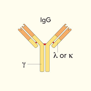
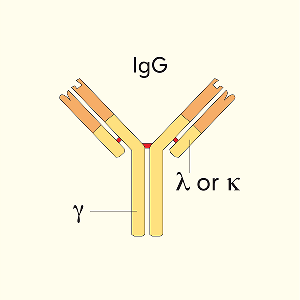
Syd Labs生产的体内实验级重组抗小鼠CD4(又称L3T4/Ly-4)单克隆抗体,克隆号 GK1.5,恒定区为小鼠IgG2b kappa(mIgG2bκ),在哺乳动物细胞中重组表达。GK1.5是小鼠免疫学研究中最经典、引用最广泛的CD4克隆之一,最早由Dialynas等人于1983年在Journal of Immunology发表,特异性识别小鼠CD4——一种主要表达在胸腺细胞(约80%)、成熟MHC II限制性辅助性T细胞(Th1/Th2/Th17/Tfh)、调节性T细胞(Treg)及部分NK-T细胞、树突状细胞上的55 kDa I型跨膜糖蛋白,是TCR共受体,通过结合MHC II类分子并招募lck激酶参与T细胞活化与胸腺分化。GK1.5可阻断MHC II–CD4相互作用,在小鼠体内经腹腔/静脉注射(通常100–500 μg/只)可有效耗竭外周与组织CD4+ T细胞,是研究肿瘤免疫、感染免疫、自身免疫病、移植耐受、疫苗应答中CD4+ T细胞功能”金标准”工具。本品为Syd Labs重组表达版本(区别于传统杂交瘤制备、带大鼠IgG2b恒定区的PA007200.r2b),恒定区为小鼠IgG2b kappa,特别适用于以小鼠同种型为背景的同型匹配对照、避免抗大鼠IgG异种反应的体内模型、以及与人/小鼠IgG Fc受体相互作用研究;经Protein A/G亲和纯化、SDS-PAGE与HPLC双检纯度>95%、LAL法内毒素<1 EU/mg、0.2 μm无菌过滤、1× PBS(pH 7.4)配方,无稳定剂无防腐剂,适合体内动物实验和各种体外功能检测。
产品参数
| 货号 | PA007200.m2b |
|---|---|
| 产品名称 | 重组抗小鼠CD4单抗(GK1.5) | Syd Labs PA007200.m2b |
| 英文名 | In vivo Grade Recombinant Anti-mouse CD4 Monoclonal Antibody, Mouse IgG2b Kappa (Clone: GK1.5) |
| 供货商名称 | Syd Labs, Inc. |
| 品牌名 | Syd Labs |
| 克隆号 | GK1.5 |
| 同种型 | 小鼠IgG2b kappa |
| 特异性 | 小鼠CD4(L3T4/Ly-4/T4),55 kDa I型跨膜糖蛋白 |
| 抗体形式 | 0.2 μM过滤的1× PBS(pH 7.4)溶液,不含稳定剂与防腐剂 |
| 内毒素 | <1 EU/mg(LAL 法测定) |
| 纯度 | >95%(还原性SDS-PAGE及HPLC检测) |
| 运输 | 本产品采用冰袋运输。收到后,请立即按下文推荐温度保存。 |
| 稳定性与存储 | 使用手动除霜冰箱,避免反复冻融;2–8 ℃可保存1个月;−20 ℃至−70 ℃可保存12个月(按供货状态) |
| 注意事项 | 仅供科研使用(For Research Use Only),不可用于人体或动物诊断与治疗;体内使用前请根据实验模型优化给药剂量与时程;长期储存建议分装避免反复冻融 |
| 产品咨询 | Syd Labs在国内只通过代理商销售其产品,不做直销。终端用户咨询价格请联系Syd Labs中国代理商。 关于Syd Labs产品如果有任何技术或其它问题,欢迎随时联系Syd Labs国内市场推广合作伙伴:武汉多找找科技有限公司,企业微信:duozhaozhao2024 联系电话:18162581039(龙经理) |
| 应用详情 | ELISA、中和实验、体内CD4+ T细胞耗竭、流式细胞术(FC/FACS)、Western Blot(WB)、免疫沉淀(IP)、免疫组化(IHC-P/IHC-F)、生物分析PK/ADA等功能实验 |
文献
体内实验级重组抗小鼠CD4单克隆抗体(克隆号GK1.5),小鼠IgG2b Kappa(In vivo Grade Recombinant Anti-mouse CD4 Monoclonal Antibody, Mouse IgG2b Kappa (Clone: GK1.5))
重组抗小鼠CD4单抗(GK1.5)背景知识:
CD4(Cluster of Differentiation 4,小鼠又称L3T4/Ly-4)是免疫球蛋白超家族成员的55 kDa I型跨膜糖蛋白,主要表达于胸腺细胞、MHC II类分子限制性的成熟辅助性T细胞(Th1/Th2/Th17/Tfh)、调节性T细胞(Treg)及部分NK-T细胞、树突状细胞和巨噬细胞表面。CD4作为T细胞受体(TCR)的共受体,通过其膜外D1/D2结构域与抗原呈递细胞表面MHC II类分子的β2结构域结合,同时其胞质尾段与蛋白酪氨酸激酶Lck相连,共同启动下游TCR信号级联,在T细胞活化、胸腺阳性/阴性选择和外周免疫应答中发挥核心作用。
GK1.5克隆是1983年由Dialynas、Wilde、Marrack、Kappler、Fitch等团队在Journal of Immunology同期发表的两篇里程碑论文中首次鉴定的大鼠抗小鼠L3T4单克隆抗体,杂交瘤由LEW大鼠经小鼠CTL克隆V4免疫后获得,抗体在SDS-PAGE还原条件下识别约52 kDa条带,表达于~80%胸腺细胞、~20%脾细胞,且与MHC II类抗原反应性高度相关。GK1.5能够深度阻断II类MHC限制性辅助T细胞的抗原特异性增殖、淋巴因子释放和抗原结合,并能在致死性打击之前阻断II类MHC反应性CTL克隆的抗原特异性杀伤——这些结果直接证明L3T4(即小鼠CD4)与人Leu-3/T4(人CD4)同源,确立了CD4作为MHC II共受体的分子本质,为后来HIV以CD4为主要受体、以及CD4+ T细胞分类和功能研究奠定了基础。
重组抗小鼠CD4单抗(GK1.5)部分参考文献:
1.Characterization of the murine T cell surface molecule, designated L3T4, identified by monoclonal antibody GK1.5: similarity of L3T4 to the human Leu-3/T4 molecule
Demetri P. Dialynas, Zang S. Quan, Katherine A. Wall, Angelica Pierres, Jose Quintans, Michael R. Loken, Michel Pierres, Frank W. Fitch
J Immunol. 1983;131(5):2445–2451. PMID: 6415170
“Monoclonal antibody GK1.5 recognizes a previously undescribed murine T cell surface molecule, designated L3T4, which migrates on SDS-PAGE under reducing conditions as a single band with an apparent m.w. of 52,000. L3T4 is expressed by approximately 80% of thymocytes and by approximately 20% of spleen cells. There appears to be poor correlation between expression of L3T4 by functional T cell clones and expression of Lyt-2, expression of the cytolytic phenotype, and class I MHC antigen reactivity. On the other hand, both a class II MHC antigen-reactive HTL clone and an Lyt-1- Mls-reactive HTL clone express L3T4. Analysis of the effect of mAb GK1.5 on PFC responses in adoptive transfer suggests that L3T4 is expressed by the helper/inducer subset of murine T cells. … mAb GK1.5 profoundly blocks antigen-specific cytolysis by the cloned class II MHC antigen-reactive CTL line A15-1.17. Antigen-specific cytolysis by A15-1.17 is blocked by mAb GK1.5 at a step before the lethal hit. Collectively, the flow cytometric, functional, and biochemical data indicate that L3T4 is similar to the human Leu-3/T4 molecule.”
2.Evidence implicating L3T4 in class II MHC antigen reactivity; monoclonal antibody GK1.5 (anti-L3T4a) blocks class II MHC antigen-specific proliferation, release of lymphokines, and binding by cloned murine helper T lymphocyte lines
David B. Wilde, Philippa Marrack, John Kappler, Demetri P. Dialynas, Frank W. Fitch
J Immunol. 1983;131(5):2178–2183. PMID: 6195255
“Monoclonal antibody GK1.5 recognizes a determinant, designated L3T4a, on the murine T cell surface molecule L3T4. The expression of L3T4a by functional murine T cell clones appears to correlate primarily with class II MHC antigen reactivity rather than with functional phenotype. … In the present studies, we demonstrate that mAb GK1.5 profoundly blocks antigen-specific proliferation and release of lymphokines by cloned murine class II MHC antigen-reactive helper T lymphocyte (HTL) lines. … Finally, we present evidence that mAb GK1.5 blocks class II MHC antigen-specific function by blocking class II MHC antigen-specific binding. The data presented here lend considerable support to the concept both that L3T4 and the human Leu-3/T4 molecules are similar and that L3T4 plays a role in class II MHC antigen-reactivity by murine T cells.”
3.CD4 T cell depletion increases memory differentiation of endogenous and CAR T cells and enhances the efficacy of Super2 and IL-33-armored CAR T cells against solid tumors
Ahmed O. Mohamed, John V. Maryanski, Anitha K. Thomas, Alex S. Gartrell-Corrado, Alex N. Salazar, Alisha N. Cash, Jeremy A. Sullivan, Yiping Yang, Gregory B. Lesinski, Jonathan H. Esensten, Surojit Sarkar
J Immunother Cancer. 2025;13(2):e009994. PMCID: PMC11815418 PMID: 39933839 DOI: 10.1136/jitc-2024-009994
“Background: The lack of durable antitumor responses by chimeric antigen receptor (CAR) T cells against solid tumors remains a major clinical hurdle. … For CD4 depletion therapy, mice were injected intraperitoneally with 250 µg per dose of anti-mouse CD4 antibody (mAb clone GK1.5; BioXcell Cat: #BE0003-1) on days 5 and 11 after tumor inoculation. … Our results demonstrate that strategic CD4 depletion can remodel the TME and enhance CAR T cell functional persistence and memory differentiation, thus representing a translatable adjunct strategy to potentiate CAR T therapy against solid tumors.”
4.MHC-II presentation by oral Langerhans cells impacts intraepithelial Tc17 abundance and Candida albicans oral infection via CD4 T cells
Peter D. Bittner-Eddy, Lori A. Fischer, Praveen Venkata Parachuru, Massimo Costalonga
Front Oral Health. 2024;5:1408255. PMCID: PMC11169704 PMID: 38871234 DOI: 10.3389/froh.2024.1408255
“In a murine model (LCΔMHC-II) designed to abolish MHC-II expression in Langerhans cells (LCs), ∼18% of oral LCs retain MHC-II, yet oral mucosal CD4 T cells numbers are unaffected. In LCΔMHC-II mice, we now show that oral intraepithelial conventional CD8αβ T cell numbers expand 30-fold. Antibody-mediated ablation of CD4 T cells in wild-type mice also resulted in CD8αβ T cell expansion in the oral mucosa. Therefore, we hypothesize that MHC class II molecules uniquely expressed on Langerhans cells mediate the suppression of intraepithelial resident-memory CD8 T cell numbers via a CD4 T cell-dependent mechanism. … Although IL-17A-expressing γδ T cells were reduced by 30%, LCΔMHC-II mice displayed greater resistance to Candida albicans in early stages of oral infection.”
5.Depleting anti-CD4 monoclonal antibody (GK1.5) treatment: influence on regulatory CD4+CD25+Foxp3+ T cells in mice
Huanfa Yi, Yu Zhen, Chun Zeng, Lianjun Zhang, Yong Zhao
Transplantation. 2008;85(7):1035–1041. PMID: 18431238 DOI: 10.1097/TP.0b013e31816a1242
“Background: CD4(+)CD25(+) regulatory T (Treg) cells are often essential for the maintenance of immunologic self-tolerance and transplant tolerance in some cases. The effects of depleting anti-CD4 monoclonal antibody (GK1.5), which was used in transplant tolerance induction, on CD4(+)CD25(+) Treg cells have not been investigated. … Results: The numbers of CD4 and CD4(+)CD25(+) Treg cells in the periphery were markedly decreased in GK1.5-treated mice. However, GK1.5 treatment significantly enhanced the ratios of CD4(+)CD25(+) T cells or CD4(+)CD25(+)Foxp3 T cells to CD4(+) T cells in the periphery (P<0.01). Compared with the control mice, more CD4(+)CD25(+) T cells in GK1.5-treated mice showed CD45RB(low) and CD62L(high) phenotype. Furthermore, enriched CD4(+)CD25(+) Treg cells in GK1.5-treated mice show immunosuppressive ability on the immune response of T effector cells to alloantigens or mitogen as efficiently as those from the control mice in vitro. Conclusions: GK1.5 could significantly enhance the percentage of CD4(+)CD25(+)Foxp3(+) Treg cells in the periphery while keeping these cells functional.”