重组抗HIV V2顶点单克隆抗体(PGT145) 人IgG1 Kappa | Syd Labs PA007184
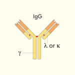
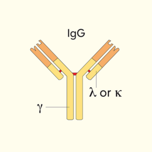
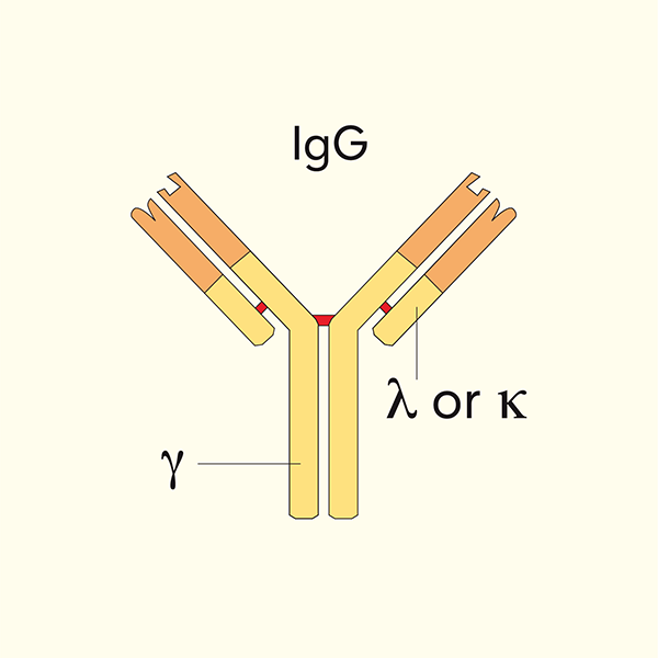
Syd Labs生产的体内实验级重组抗HIV V2顶点单克隆抗体(克隆PGT145)为哺乳动物细胞表达的全长人IgG1 Kappa抗体,是Walker/Burton团队2011年从非洲精英中和者(IAVI Protocol G Donor 84)中与PGT141-PGT144一同分离的V2顶端(V2-apex)广谱中和抗体代表成员;重链使用IGHV1-8*02胚系基因,携带约33个氨基酸、含双硫酸化酪氨酸(Tyr33/Tyr34)的超长CDRH3,以近似垂直于三聚体顶端的角度结合融合前闭合态Env三聚体V2顶点N160糖基超位点(四元表位,quaternary epitope),识别V2 β-桶C链及N160/N156/N173高甘露糖簇,对约78%跨亚型HIV-1假病毒具高中和活性(几何平均IC50约0.29 μg/mL);PGT145最广泛的应用是作为亲和层析配基(PGT145 affinity column)从细胞上清中一步纯化天然闭合态SOSIP/Env三聚体,是HIV结构生物学与疫苗领域纯化native-like Env trimers的金标准工具抗体,同时适用于HIV-1中和实验、Env三聚体顶端结构生物学、V2顶点糖基识别机制研究、V1V2疫苗抗原性评价及多种体内外功能实验。
产品参数
| 货号 | PA007184 |
|---|---|
| 产品名称 | 重组抗HIV V2顶点单克隆抗体(PGT145) 人IgG1 Kappa | Syd Labs PA007184 |
| 英文名 | In Vivo Grade Recombinant Anti-HIV V2 Apex Monoclonal Antibody (Clone PGT145), Human IgG1 Kappa |
| 供货商名称 | Syd Labs, Inc. |
| 品牌名 | Syd Labs |
| 克隆号 | PGT145 |
| 同种型 | 人 IgG1,Kappa |
| 反应性 | HIV-1(跨亚型,约 78% 假病毒被中和,几何平均 IC50 约 0.29 μg/mL;严格依赖N160糖基与三聚体闭合态构象) |
| 特异性 | HIV gp120 V2顶点(V2-apex,N160糖基超位点,三聚体四元构象表位) |
| 抗体形式 | 0.2 μm 过滤的 1× PBS 溶液 |
| 内毒素 | <1 EU/mg(LAL 法测定) |
| 纯度 | >95%(还原性 SDS-PAGE) |
| 运输 | 冰袋运输,收到后请立即按推荐条件储存 |
| 稳定性与存储 | 收到后 2–8 ℃ 保存 1 个月;−20 ℃ 至 −70 ℃ 保存 12 个月;避免反复冻融,建议使用可手动除霜的冰箱 |
| 产品咨询 | Syd Labs在国内只通过代理商销售其产品,不做直销。终端用户咨询价格请联系Syd Labs中国代理商。 关于Syd Labs产品如果有任何技术或其它问题,欢迎随时联系Syd Labs国内市场推广合作伙伴:武汉多找找科技有限公司,企业微信:duozhaozhao2024 联系电话:18162581039(龙经理) |
| 应用详情 | ELISA、Western blot、免疫组化(IHC)、流式细胞术(FC)、HIV-1中和实验、gp120外域/CD4bs识别机制研究、Env结构生物学(结晶辅助与抗原性分析)、抗体逃逸通路分析及多种体内外功能实验 |
文献
重组抗HIV V2顶点单克隆抗体(PGT145) 人IgG1 Kappa,体内实验级(In Vivo Grade Recombinant Anti-HIV V2 Apex Monoclonal Antibody (Clone PGT145), Human IgG1 Kappa)
重组抗HIV V2顶点单克隆抗体背景知识:
HIV-1 Env三聚体顶端(apex)由三个gp120亚基的V1V2域环抱构成,是病毒与宿主共受体相互作用、构象重排启动以及广谱中和抗体靶向的关键区域。V2顶点(V2-apex)特指以V2 β-桶C链为中心、N160高甘露糖为核心糖基的超位点(N160-glycan supersite),被PG9/PG16、PGT145、CH01–CH04、PGT141–145、CAP256-VRC26等多家族bNAb共同靶向。这些抗体均具有三聚体特异性(quaternary-specific)——仅结合天然闭合态三聚体而不识别单体gp120或开放态Env——并以超长CDRH3穿透糖基屏障,以硫酸化酪氨酸接触N160糖基、通过链-链β-氢键结合V2 C链,实现高效中和。
克隆PGT145由Burton团队2011年在Nature上报道,与PGT141–PGT144同属从精英中和者IAVI Protocol G Donor 84分离的”第二代”V2-apex bNAb。与PG9相比,PGT145重链使用不同的VH胚系(IGHV1-802而非IGHV3-3301),CDRH3更长(约33 aa)且带有两个硫酸化酪氨酸(Tyr100h与Tyr100i),以更垂直于三聚体顶端的角度接近V2 C链,接触N160、N156、N173等糖基及V2残基Lys168/Tyr173/Asn160等,在162株跨亚型假病毒组中约78%被中和,几何平均IC50约0.29 μg/mL,且具有较低的多反应性(non-polyreactive),适合体内应用。
PGT145在HIV Env结构生物学和疫苗领域最核心、最不可替代的应用是作为亲和层析配基(PGT145 affinity column):2015年Pugach等在Nature Communications报道了基于PGT145 IgG偶联树脂的亲和层析方法,可以从BG505 SOSIP.664、B41 SOSIP.664、JR-FL SOSIP等多种亚型SOSIP免疫原转染细胞上清中,一步、高效、非变性地纯化出闭合态、高纯度、抗原性完整的native-like Env三聚体,同时有效分离出单体、二聚体、开放态等杂质。该方法迅速成为HIV结构生物学和疫苗领域制备高纯度Env三聚体抗原的金标准——几乎所有后续的SOSIP高分辨冷冻电镜结构、V1V2疫苗免疫原制剂、bNAb共结晶样品均以PGT145亲和柱作为核心纯化步骤。2017年Lee等在Nature Structural & Molecular Biology上发表的PGT145 Fab与完整膜提取Env三聚体复合物的冷冻电镜结构,进一步在天然膜环境中验证了PGT145的结合角度与识别机制;Gorman等2016年的V1V2-Fab共晶研究也将PGT145纳入与PG9、PG16、CH03等V2-apex抗体的系统性结构比较,为V2靶向疫苗设计提供了共同结构模板。
本品为体内实验级(In Vivo Grade)制剂,来源于哺乳动物细胞表达的重组全长人IgG1 Kappa,内毒素低于1 EU/mg、配方为无防腐剂的1× PBS,既适合用于HIV-1中和实验、Env三聚体顶端V2 N160糖基超位点识别机制研究、SOSIP亲和柱制备(可偶联至CNBr-或NHS-activated Sepharose)、Env三聚体冷冻电镜/结晶样品制备与抗原性分析、V1V2靶向疫苗免疫原质量评价,也可直接用于SHIV恒河猴/人源化小鼠体内被动保护或抗体治疗研究等需要低内毒素背景的功能实验。使用前请根据实验体系确定工作浓度,并避免反复冻融;用于亲和柱偶联时建议按0.5–2 mg IgG/mL树脂比例进行偶联。
重组抗HIV V2顶点单克隆抗体部分参考文献:
1.Broad neutralization coverage of HIV by multiple highly potent antibodies Laura M Walker,et al.Nature. 2011.PMCID: PMC3177487 “Broadly neutralizing antibodies (bNAbs) to HIV-1 are of great importance for vaccine design and immunotherapy. Here we describe the isolation of 290 new anti-HIV-1 monoclonal antibodies from the memory B cells of four donors selected for exceptional plasma neutralization breadth and potency. Among these, we identify a new family of bNAbs (PGT141-PGT145) that targets a quaternary glycan-dependent epitope in the V2 region of gp120 at the trimer apex, related to but distinct from the previously described PG9/PG16 epitope. PGT145 neutralized 78% of 162 cross-clade pseudoviruses with a geometric mean IC50 of 0.29 µg/ml and used the IGHV1-8 germline gene with a remarkably long, 33-residue tyrosine-sulfated CDR H3. These findings expand the spectrum of HIV-1 antibodies capable of potent and broad neutralization and provide additional tools and templates for vaccine design.”
2. A native-like SOSIP.664 trimer based on an HIV-1 subtype B env gene Pavel Pugach,et al.Nat Commun. 2015.PMCID: PMC4695221 “Soluble, cleaved recombinant HIV-1 envelope glycoprotein (Env) trimers of the SOSIP.664 design are leading native-like vaccine candidates. The BG505 SOSIP.664 trimer (clade A) has been structurally and antigenically well characterized, but trimers from other clades are needed to address global sequence diversity. Here we describe the design and characterization of B41 SOSIP.664, a clade B native-like trimer derived from a transmitted/founder virus. B41 SOSIP.664 forms homogeneous native-like trimers that can be purified to near-homogeneity by a single-pass affinity chromatography on PGT145 antibody columns. Negative-stain electron microscopy and antigenic profiling with bNAbs (including PGT145, PG9, PG16, PGT128, 35O22 and VRC01) and non-NAbs confirm that PGT145-purified B41 SOSIP.664 adopts a compact, prefusion conformation closely matching the BG505 SOSIP.664 crystal structure. The PGT145 affinity-purification strategy provides a broadly applicable method to isolate native-like SOSIP trimers from diverse clades for structural and vaccine studies.”
3. Crystal structure of a soluble cleaved HIV-1 envelope trimer Jean-Philippe Julien,et al.Science. 2013.PMCID: PMC3885649 “HIV-1 entry into CD4+ T cells is mediated by the envelope (Env) glycoprotein spike, a trimer of gp120-gp41 heterodimers. Here we report the crystal structure at 4.7 Å resolution of the soluble, cleaved BG505 SOSIP.664 trimer, the first atomic-resolution structure of a native-like HIV-1 Env trimer. The trimer adopts a compact, mushroom-shaped conformation, with V1V2 and V3 loops sequestered at the trimer apex. Antigenic mapping with a panel of bNAbs shows that the trimer apex quaternary epitopes recognized by PG9, PG16 and PGT145 are well preserved, while non-neutralizing epitopes are largely occluded. The structure defines the overall architecture of the prefusion Env spike and provides a high-resolution template for structure-based HIV-1 vaccine design.”
4. Structures of HIV-1 Env V1V2 with broadly neutralizing antibodies reveal commonalities that enable vaccine design Jason Gorman,et al.Nat Struct Mol Biol. 2016.PMCID: PMC4722598 “Broadly neutralizing antibodies (bNAbs) against the V1V2-apex site of the HIV-1 Env trimer are promising targets for vaccine design. To define common structural features of V1V2 recognition, we determined crystal structures of bNAbs PG9, PG16, PGT145 and CH03 in complex with scaffolded V1V2 domains, and analyzed these together with high-resolution Env trimer structures. All V1V2-apex bNAbs use long, tyrosine-sulfated CDR H3 loops to penetrate the glycan shield and engage a conserved β-strand motif in V1V2 through strand-strand interactions involving the N160 glycan. PGT145 uses an IGHV1-8-encoded heavy chain with a bisulfated 33-residue CDR H3 that approaches V1V2 at a slightly more perpendicular angle than PG9/PG16, while maintaining the same core N160 glycan-recognition mode. These results highlight common structural requirements for potent V1V2 neutralization and provide atomic-level templates for eliciting PGT145-like bNAbs by vaccination.”
5. Broadly neutralizing anti-HIV-1 antibodies from a single donor target the V2 apex with distinct modes of recognition Jeong Hyun Lee,et al.Nat Struct Mol Biol. 2017.PMCID: PMC5449314 “V2-apex-directed bNAbs target a quaternary, glycan-dependent epitope at the HIV-1 Env trimer apex and are promising for therapy and vaccine design. Here we report the isolation and characterization of a panel of V2-apex bNAbs from donor IAVI Protocol G 84, including PGT145 and related somatic variants. Cryo-EM structures of these Fabs in complex with membrane-extracted, fully glycosylated native Env trimers reveal that PGT145 binds the trimer apex at a near-perpendicular angle, with its bisulfotyrosine-containing CDR H3 inserting deep into the glycan shield to contact N160 and the V2 C strand. Comparison with PG9 and PG16 shows that V2-apex antibodies can access the same supersite through slightly different trajectories, and viral escape from PGT145 selects for mutations that disrupt N160 glycan processing or alter V2 residues that contact the antibody. These findings provide a structural framework for understanding the development and breadth of V2-apex bNAb responses and guide V2-targeted immunogen design.”