
重组抗小鼠CD5单抗(53-7.313) | Syd Labs PA007696.r2a
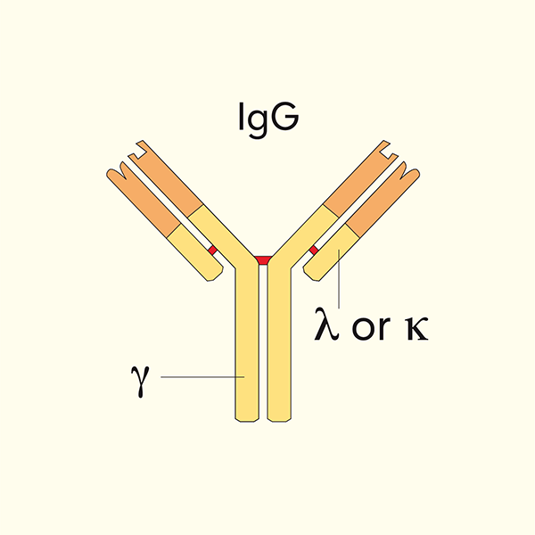
Syd Labs体内实验级重组抗小鼠 CD5 单克隆抗体,克隆号 53-7.313,恒定区为大鼠 IgG2a Kappa(Rat IgG2a Kappa),由哺乳动物细胞重组表达,靶向小鼠 CD5 分子。CD5 又称 Lyt-1(T细胞抗原Lyt-1)、Ly-1(淋巴细胞抗原1)、T1(T细胞抗原T1)、Tp67(67 kDa T细胞磷糖蛋白)、Ly-12、Ly-A,是一种约 67 kDa I 型跨膜糖蛋白(基因名 Cd5,小鼠19号染色体、人类11号染色体11q12.2),属于富含半胱氨酸清道夫受体超家族(SRCRSF,scavenger receptor cysteine-rich superfamily),胞外从 N 端依次含 3 个 SRCR(清道夫受体富半胱氨酸)结构域,无 Ig 样结构域;胞浆区长尾含一个免疫受体酪氨酸抑制基序样基序(ITIM-like motif),磷酸化后可招募酪氨酸磷酸酶 SHP-1,对 TCR/BCR 信号传递起负调控作用。CD5 在几乎所有胸腺细胞和成熟 T 细胞表面高组成性表达,在小鼠 B-1a 细胞(腹腔 CD5⁺ B 细胞)上低表达,是鉴定 T 细胞谱系与胸腺细胞分化阶段最经典的标志之一。CD5 通过与配体 CD72(B 细胞表面)及其他尚未完全阐明的配体结合,调控 T 细胞活化阈值、胸腺正负选择、B1 细胞发育、外周免疫耐受,并参与病原体相关分子模式(PAMP)识别与 IL-10 分泌调节。克隆 53-7.313 是 Ledbetter 与 Herzenberg 于 1979 年建立的经典大鼠抗小鼠 Lyt-1/CD5 单克隆抗体(大鼠 IgG2aκ,53-7.313 为原杂交瘤亚克隆编号),是最早用于鉴定小鼠 T 细胞亚群(Lyt-1⁺2⁻ vs Lyt-1⁺2⁺)的标志性抗体之一,广泛用于流式细胞术鉴定 T 细胞/B1 细胞、T 细胞活化/增殖调节、免疫细胞亚群分群、中和/功能实验,以及 CD5 信号通路在免疫耐受、自身免疫病、白血病、感染模型中的研究。本品经 Protein G 亲和纯化,还原性 SDS-PAGE 及 HPLC 检测纯度>95%,LAL 法内毒素<1 EU/mg,0.2 μM 过滤于 pH 7.4、不含稳定剂或防腐剂的 1× PBS 溶液,适用于 ELISA、中和实验、功能实验(如生物分析 PK 与 ADA 检测)以及研究受小鼠 CD5 蛋白影响的生物学通路的实验和体内动物实验。
产品参数
| 货号 | PA007696.r2a |
|---|---|
| 产品名称 | 重组抗小鼠CD5单抗(53-7.313) | Syd Labs PA007696.r2a |
| 英文名 | In Vivo Grade Recombinant Anti-mouse CD5 Monoclonal Antibody (Clone: 53-7.313), Rat IgG2a Kappa |
| 供货商名称 | Syd Labs, Inc. |
| 品牌名 | Syd Labs |
| 别称 | Lyt-1(T细胞抗原Lyt-1)、Ly-1(淋巴细胞抗原-1/Lymphocyte antigen-1)、T1(T细胞抗原T1)、Tp67(67 kDa T细胞磷糖蛋白)、Ly-12、Ly-A |
| 克隆号 | 53-7.313 |
| 同种型 | 大鼠 IgG2a Kappa |
| 特异性 | 该体内实验级重组大鼠单克隆抗体(克隆号 53-7.313)特异性结合小鼠 CD5 |
| 抗体形式 | 0.2 μM 过滤的溶液,pH 7.4,不含稳定剂或防腐剂 |
| 内毒素 | LAL 法测定,低于每 1 mg 蛋白 1 EU |
| 纯度 | 还原性条件下 SDS-PAGE 及 HPLC 检测>95% |
| 运输 | 采用冰袋运输;收到后请立即按下方推荐温度保存 |
| 稳定性与存储 | 使用手动除霜冰箱,避免反复冻融;自收货之日起 12 个月,按供货状态于 −20 ℃ 至 −70 ℃ 保存;自收货之日起 1 个月,按供货状态于 2 ℃ 至 8 ℃ 保存 |
| 产品咨询 | Syd Labs在国内只通过代理商销售其产品,不做直销。终端用户咨询价格请联系Syd Labs中国代理商。 关于Syd Labs产品如果有任何技术或其它问题,欢迎随时联系Syd Labs国内市场推广合作伙伴:武汉多找找科技有限公司,企业微信:duozhaozhao2024 联系电话:18162581039(龙经理) |
| 应用详情 | ELISA、中和实验、功能实验(如生物分析 PK 与 ADA 检测),以及研究受小鼠 CD5 蛋白影响的生物学通路的实验 |
文献
PA007696.r2a: Syd Labs 重组抗小鼠CD5单克隆抗体(克隆号53-7.313) ,体内实验级重组,大鼠IgG2a Kappa(In Vivo Grade Recombinant Anti-mouse CD5 Monoclonal Antibody (Clone: 53-7.313), Rat IgG2a Kappa)
重组抗小鼠CD5单抗(53-7.313)背景知识:
CD5 是一种在 T/B 淋巴细胞谱系表达的跨膜糖蛋白,也是小鼠免疫学研究史上最早被单克隆抗体鉴定的 T 细胞分化抗原之一。其多个别名反映了不同实验室独立发现与命名历史:
Lyt-1:20 世纪 70 年代通过异种血清(alloantisera)鉴定的小鼠 T 细胞 Lyt 抗原系列之一,最初定义”辅助性 T 细胞”表型为 Lyt-1⁺2⁻,是小鼠 T 细胞亚群研究的第一个标志系统;
Ly-1(Lymphocyte antigen-1):小鼠 Ly 抗原编号体系;
T1 / Tp67:人类 CD5 最初被称为 T1(Leu-1),因 SDS-PAGE 分子量约 67 kDa 又称 Tp67(T 细胞 67 kDa 磷蛋白);
Ly-12 / Ly-A:早期小鼠遗传学研究中的等位基因命名。
CD5 为约 67 kDa I 型跨膜糖蛋白,属于 SRCR 超家族(清道夫受体富半胱氨酸超家族,与 CD6、M130/CD163、Spα、DMBT1 同家族),胞外区从 N 端依次为 3 个 SRCR 结构域(D1–D3),不含 Ig 样结构域,D1 域含 CD72 等配体结合位点;跨膜区后是一段较长的胞浆尾,含一个膜近端 ITIM-like 基序(Y378 位点,小鼠/人保守)以及多个可被 Lck、Fyn 等 Src 家族激酶磷酸化的酪氨酸,磷酸化后可招募 SHP-1、SHP-2 及 Cbl 等信号负调节分子。
重组抗小鼠CD5单抗(53-7.313)部分参考文献:
1. Xenogeneic monoclonal antibodies to mouse lymphoid differentiation antigens.
Ledbetter JA, Herzenberg LA
Immunol Rev. 1979;47:63–90. PMID: 91593 DOI: 10.1111/j.1600-065x.1979.tb00283.x
“Xenogeneic rat monoclonal antibodies directed against several mouse lymphocyte differentiation antigens have been prepared by fusing rat immune spleen cells with the mouse myeloma line NS-1. The rat antibodies were specific for Thy-1.2, Lyt-1.2, Lyt-2.2 and Lyt-3.1, as shown by cytotoxicity testing and immunoprecipitation studies. The Lyt-1-specific monoclonal antibody (clone 53-7.3, subclone 53-7.313) precipitated a polypeptide of approximately 67,000 daltons (Lyt-1/Tp67) from T cells. These antibodies have been used to define and separate mouse T cell subsets by flow cytometry and complement-mediated cytotoxicity. The results establish Lyt-1 as a pan-T marker present on all peripheral T cells, and Lyt-2 and Lyt-3 as markers of the cytotoxic/suppressor T cell subset, providing the basis for modern T lymphocyte subset analysis.”
2.T cell subsets defined by expression of Lyt-1,2,3 and Thy-1 antigens. Two-parameter immunofluorescence and cytotoxicity analysis with monoclonal antibodies modifies current views.
Ledbetter JA, Rouse RV, Micklem HS, Herzenberg LA
J Exp Med. 1980 Aug 1;152(2):280–295. PMID: 6967469 PMCID: PMC2185929 DOI: 10.1084/jem.152.2.280
“Using monoclonal antibodies and two-color flow cytometry as well as cytotoxicity assays, we have re-examined the distribution of Lyt antigens on mouse T cells. In contrast to the previously held view that mature peripheral T cells fall into two mutually exclusive subpopulations—Lyt-1⁺2⁻ helper cells and Lyt-1⁻2⁺ cytotoxic/suppressor cells—we find that essentially all mature peripheral T cells express Lyt-1, while Lyt-2 and Lyt-3 are expressed together on a subset (approximately 30–40% of peripheral T cells, the cytotoxic/suppressor lineage). A small Lyt-1⁻2⁺ population (less than 5%) in spleen appears to represent immature thymocytes or a minor non-T population. These findings revise the T cell subset model and indicate that Lyt-1 is a pan-T marker whose expression is necessary but not sufficient to define helper T cells, and that Lyt-2/3 expression identifies the class I MHC–restricted cytotoxic/suppressor subset. Monoclonal antibody 53-7.313 was used throughout these studies as the anti-Lyt-1 reagent.”
3.Effects of Lyt antibodies on T-cell functions: Augmentation by anti-Lyt-1 as opposed to inhibition by anti-Lyt-2.
Hollander N, Pillemer E, Weissman IL
Proc Natl Acad Sci U S A. 1981 Feb;78(2):1148–1151. PMID: 6970765 PMCID: PMC319966 DOI: 10.1073/pnas.78.2.1148
“Monoclonal anti-Lyt-1 (53-7.313) and anti-Lyt-2 manifest inverse effects on allogeneic mixed lymphocyte reactions when added to the reaction mixtures without complement. Anti-Lyt-1 augments cell proliferation and generation of cytotoxic T cells but has no effect on cell-mediated cytolysis, whereas anti-Lyt-2 blocks cell proliferation, generation of killer cells, and cytolytic activity of killer cells. The augmenting effect of anti-Lyt-1 cannot be attributed to a direct mitogenic effect on T cells. Both inhibition by anti-Lyt-2 and potentiation by anti-Lyt-1 require interaction of responder cells with the antibodies during the first 24 hr of the mixed lymphocyte reaction, indicating that early stages of the reaction are sensitive to Lyt antibodies. The enhancing effect of anti-Lyt-1 on alloantigen-induced T-cell proliferation is associated with augmented production of T-cell growth factors (interleukin-2) and interferon. These results provide the first evidence that the Lyt-1 molecule is functionally involved in T cell activation and lymphokine production.”
4.CD5 negatively regulates the T-cell antigen receptor signal transduction pathway: involvement of SH2-containing phosphotyrosine phosphatase SHP-1.
Perez-Villar JJ, Whitney GS, Bowen MA, Hewgill DH, Aruffo AA, Kanner SB
Mol Cell Biol. 1999 Apr;19(4):2903–2912. PMCID: PMC84084 PMID: 10082557 DOI: 10.1128/MCB.19.4.2903
“The negative regulation of T- or B-cell antigen receptor signaling by CD5 was proposed based on studies of thymocytes and peritoneal B-1a cells from CD5-deficient mice. Here, we show that CD5 is constitutively associated with phosphotyrosine phosphatase activity in Jurkat T cells. CD5 was found associated with the Src homology 2 (SH2) domain containing hematopoietic phosphotyrosine phosphatase SHP-1 in both Jurkat cells and normal phytohemagglutinin-expanded T lymphoblasts. This interaction was increased upon T-cell receptor (TCR)-CD3 cell stimulation. CD5 co-cross-linking with the TCR-CD3 complex down-regulated the TCR-CD3-increased Ca2+ mobilization in Jurkat cells. In addition, stimulation of Jurkat cells or normal phytohemagglutinin-expanded T lymphoblasts through TCR-CD3 induced rapid tyrosine phosphorylation of several protein substrates, which was substantially diminished after CD5 cross-linking. The CD5-regulated substrates included CD3ζ, ZAP-70, Syk, and phospholipase Cγ1 but not the Src family tyrosine kinase p56lck. By mutation of all four CD5 intracellular tyrosine residues to phenylalanine, we found the membrane-proximal tyrosine at position 378, which is located in an immunoreceptor tyrosine-based inhibitory (ITIM)-like motif, crucial for SHP-1 association. The F378 point mutation ablated both SHP-1 binding and the down-regulating activity of CD5 during TCR-CD3 stimulation. These results suggest a critical role of the CD5 ITIM-like motif, which by binding to SHP-1 mediates the down-regulatory activity of this receptor.”
5.The immunomodulatory properties of the CD5 lymphocyte receptor in health and disease.
Soldevila G, Raman C, Lozano F
Immunol Rev. 2011 May;241(1):126–142. PMCID: PMC3109098 PMID: 21482089 DOI: 10.1111/j.1600-065X.2011.01011.x
“CD5 is a scavenger-like receptor expressed in association with the antigen-specific receptors on T and B-1a lymphocytes. Recent studies reveal a broader biology for CD5 that includes its role as regulator of cell death and as a receptor for pathogen-associated molecular patterns, in addition to its previously described function as an inhibitory receptor. These findings shed new light into the mechanistic role of CD5 in leukemias and effector cells to exogenous (infectious) or endogenous (autoimmune, tumoral) antigens. The newly identified properties make this receptor a potential candidate to be targeted for therapeutic intervention as well as immune modulation. This review describes the current knowledge on the function of CD5 as an immunomodulatory receptor both in health and disease.”