
定点生物素化重组抗HA标签单抗(克隆号12CA5),人IgG1 Fc Silent Kappa | Syd Labs PA006804.h1Fs-b
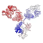

定点生物素化重组抗HA标签单抗(克隆号12CA5)是由哺乳动物细胞表达、经重链C端Avi标签定点生物素化的人IgG1 Fc Silent Kappa嵌合抗体,由小鼠可变区与人IgG1 kappa恒定区组成。其克隆12CA5源自抗流感病毒血凝素(HA)表位YPYDVPDYA的单克隆抗体,可识别HA融合蛋白上N端、Met N端、C端及内部位点的表位。每个抗体分子通过定点生物素化携带一个生物素,与链霉亲和素/亲和素系统配合时信号均一、灵敏度高,适用于Western Blot、免疫沉淀、免疫荧光、免疫组化、流式细胞术、染色质免疫沉淀等实验;Fc段经沉默改造,可减少与Fc受体、补体及蛋白A/G的非特异性结合,降低背景。
产品参数
| 货号 | PA006804.h1Fs-b |
|---|---|
| 产品名称 | 定点生物素化重组抗HA标签单抗(克隆号12CA5),人IgG1 Fc Silent Kappa | Syd Labs PA006804.h1Fs-b |
| 英文名 | Biotinylated Recombinant Anti-HA-Tag Monoclonal Antibody (Clone 12CA5), Human IgG1 Fc Silent Kappa |
| 供货商名称 | Syd Labs, Inc. |
| 品牌名 | Syd Labs |
| 别称 | 抗HA抗体、抗HA标签抗体、抗HA-Tag单抗(12CA5)、生物素化抗HA抗体、HA.11 |
| 概述 | 定点生物素化重组抗HA标签人IgG1单抗(克隆号12CA5),经Avi标签在重链C端定点标记生物素,识别YPYDVPDYA表位,Fc段经沉默改造以降低背景 |
| 克隆号 | 12CA5 |
| 同种型 | 人 IgG1 Fc Silent Kappa |
| 反应性 | 人、小鼠、大鼠、其它动物 |
| 特异性 | 检测和纯化HA融合蛋白,无论HA序列是否存在于融合蛋白的N-末端、Met N-末端、C-末端或内部位点;识别流感病毒血凝素YPYDVPDYA表位 |
| 应用 | ELISA、流式细胞术、免疫印迹、免疫沉淀、免疫细胞化学、免疫荧光、染色质免疫沉淀、电子显微镜、超位移测定、与琼脂糖或磁珠结合的蛋白质纯化以及同型对照 |
| 纯化 | 采用蛋白A亲和层析法,从化学成分明确的培养基中培养的哺乳动物细胞上清液中亲和纯化 |
| 抗体形式 | 0.2 μm过滤的1×PBS溶液 |
| 纯度 | >95%(还原条件下SDS-PAGE及HPLC测定) |
| 运输 | 采用冰袋运输。收到后请立即按建议温度储存 |
| 稳定性与存储 | 使用手动除霜冰箱并避免反复冻融循环。2-8°C自收到之日起可保存1个月;-20至-70°C自收到之日起可保存12个月 |
| 注意事项 | PA006804.h1Fs-b Syd Labs:该抗体为定点生物素化的重组抗HA标签人IgG1单抗(克隆号12CA5),Fc Silent设计可减少与Fc受体、补体及蛋白A/G的非特异结合;定点生物素化保证每分子抗体携带一个生物素,与链霉亲和素系统配合时信号均一、灵敏度高。批量订购价格更低。 |
| 产品咨询 | Syd Labs在国内只通过代理商销售其产品,不做直销。终端用户咨询价格请联系Syd Labs中国代理商。 关于Syd Labs产品如果有任何技术或其它问题,欢迎随时联系Syd Labs国内市场推广合作伙伴:武汉多找找科技有限公司,企业微信:duozhaozhao2024 联系电话:18162581039(龙经理) |
文献
PA006804.h1Fs-b: Syd Labs定点生物素化重组抗HA标签单抗(克隆号12CA5),人IgG1 Fc Silent Kappa(Biotinylated Recombinant Anti-HA-Tag Monoclonal Antibody (Clone 12CA5), Human IgG1 Fc Silent Kappa)
重组抗HA标签抗体(克隆号12CA5)部分参考文献:
1. Olfactory inputs to appetite neurons in the hypothalamus Donghui Kuang,et al.Proc Natl Acad Sci U S A. 2026. PMID: 41591908 “The sense of smell has potent effects on appetite, but the underlying neural pathways remain undefined. Here, we investigated how olfactory signals reach two subsets of appetite-linked (“appetite”) neurons in the hypothalamic arcuate nucleus: Agouti-related peptide (AgRP) neurons, which stimulate appetite, and POMC (pro-opiomelanocortin) neurons, which suppress it. Using polysynaptic viral tracing, we show that AgRP and POMC neurons receive indirect input from partially overlapping but distinct areas of the olfactory cortex, indicating that they process different sets of olfactory information. We also identify different complements of neurons more directly upstream of AgRP and POMC neurons that could relay olfactory cortical signals to the appetite neurons. Single-cell transcriptomics shows heterogeneous expression of neuromodulator receptors among AgRP neurons.”
2. A calcium-sensing receptor dileucine motif directs internalization to spatially distinct endosomal signaling pathways Rachael A. Wyatt,et al.iScience. 2025. PMCID: PMC12159506 “G protein-coupled receptors (GPCRs) can temporally alter signaling by generating secondary signals from internalized receptors. However, the motifs required to direct GPCRs to these pathways are ill defined. The GPCR, calcium-sensing receptor (CaSR), regulates parathyroid hormone release by promiscuous G protein coupling at cell surfaces and by selectively recruiting Gq/11 to early endosomal membranes. Here, we demonstrate that a dileucine endocytic motif is required for directing CaSR to dynamic spatiotemporal pathways by interacting with the adaptor protein-2 σ-subunit, which is mutated in hypercalcemic patients. From early endosomes, internalized CaSR is targeted to spatially distinct pathways involving early-to-recycling and late endosomes, to direct compartment-specific signals and regulate GPCR forward trafficking. The clinically available allosteric modulator cinacalcet biases signaling to preferentially enhance early endosomal-to-recycling pathways, demonstrating the veracity of targeting discrete endosomal pathways. These results reveal that single GPCRs can simultaneously activate spatially distinct endosomal pathways and demonstrate the structural motifs that facilitate such multidimensional signaling.”
3. Subtype-specific conformational landscape of NMDA receptor gating Julia Bleier,et al.Cell Rep. 2024. PMCID: PMC11446236 “N-methyl-D-aspartate receptors are ionotropic glutamate receptors that mediate synaptic transmission and plasticity. Variable GluN2 subunits in diheterotetrameric receptors with identical GluN1 subunits set very different functional properties. To understand this diversity, we use single-molecule fluorescence resonance energy transfer (smFRET) to measure the conformations of the ligand binding domain and modulatory amino-terminal domain of the common GluN1 subunit in receptors with different GluN2 subunits. Our results demonstrate a strong influence of the GluN2 subunits on GluN1 rearrangements, both in non-agonized and partially agonized activation intermediates, which have been elusive to structural analysis, and in the fully liganded state. Chimeric analysis reveals structural determinants that contribute to these subtype differences. Our study provides a framework for understanding the conformational landscape that supports highly divergent levels of activity, desensitization, and agonist potency in receptors with different GluN2s and could open avenues for the development of subtype-specific modulators.”
4. Distinct beta-arrestin coupling and intracellular trafficking of metabotropic glutamate receptor homo- and heterodimers Joon Lee,et al.Sci Adv. 2023. PMCID: PMC10699790 “The metabotropic glutamate receptors (mGluRs) are family C, dimeric G protein–coupled receptors (GPCRs), which play critical roles in synaptic transmission. Despite an increasing appreciation of the molecular diversity of this family, how distinct mGluR subtypes are regulated remains poorly understood. We reveal that different group II/III mGluR subtypes show markedly different beta-arrestin (β-arr) coupling and endocytic trafficking. While mGluR2 is resistant to internalization and mGluR3 shows transient β-arr coupling, which enables endocytosis and recycling, mGluR8 and β-arr form stable complexes, which leads to efficient lysosomal targeting and degradation. Using chimeras and mutagenesis, we pinpoint carboxyl-terminal domain regions that control β-arr coupling and trafficking, including the identification of an mGluR8 splice variant with impaired internalization. We then use a battery of high-resolution fluorescence assays to find that heterodimerization further expands the diversity of mGluR regulation. Together, this work provides insight into the relationship between GPCR/β-arr complex formation and trafficking while revealing diversity and intricacy in the regulation of mGluRs.”
5. Homo- and hetero-dimeric subunit interactions set affinity and efficacy in metabotropic glutamate receptors Chris Habrian,et al.Nat Commun. 2023. PMCID: PMC10719366 “Metabotropic glutamate receptors (mGluRs) are dimeric class C G-protein-coupled receptors that operate in glia and neurons. Glutamate affinity and efficacy vary greatly between the eight mGluRs. The molecular basis of this diversity is not understood. We used single-molecule fluorescence energy transfer to monitor the structural rearrangements of activation in the mGluR ligand binding domain (LBD). In saturating glutamate, group II homodimers fully occupy the activated LBD conformation (full efficacy) but homodimers of group III mGluRs do not. Strikingly, the reduced efficacy of Group III homodimers does not arise from differences in the glutamate binding pocket but, instead, from interactions within the extracellular dimerization interface that impede active state occupancy. By contrast, the functionally boosted mGluR II/III heterodimers lack these interface ‘brakes’ to activation and heterodimer asymmetry in the flexibility of a disulfide loop connecting LBDs greatly favors occupancy of the activated conformation. Our results suggest that dimerization interface interactions generate substantial functional diversity by differentially stabilizing the activated conformation. This diversity may optimize mGluR responsiveness for the distinct spatio-temporal profiles of synaptic versus extrasynaptic glutamate.”
重组抗HA标签抗体(克隆号12CA5)背景知识
HA表位标签与12CA5克隆
HA标签源自人流感病毒血凝素(hemagglutinin,HA)蛋白的一段九肽YPYDVPDYA(约1.1 kDa),是使用最广泛的表位标签之一。12CA5是用该9肽免疫Balb/c小鼠获得的经典抗HA单克隆抗体,可识别N端、Met N端、C端及内部位点的HA表位,被大量文献用于检测与纯化HA融合蛋白。
定点生物素化与链霉亲和素系统
本产品通过抗体重链C端Avi标签实现定点生物素化,每个抗体分子携带一个生物素,避免了化学随机标记造成的活性损失与批间差异。配合链霉亲和素/亲和素偶联的酶、荧光基团或磁珠,可进行信号放大、多重检测与蛋白捕获。
Fc Silent 与嵌合人IgG1
该抗体为嵌合抗体:保留小鼠可变区(决定对HA表位的特异性),恒定区换为人IgG1 kappa。Fc Silent 指Fc段经改造后不再结合Fc受体、补体及蛋白A/G等,可显著降低在Fc受体丰富的细胞与组织样本中的非特异背景。
推荐应用场景
适合Western Blot、免疫沉淀/共沉淀、免疫荧光、免疫组化、流式细胞术、染色质免疫沉淀,以及以链霉亲和素磁珠/琼脂糖进行的HA融合蛋白纯化与拉下实验。