


重组抗生物素抗体,小鼠IgG2a Kappa | Syd Labs PA007522.m2a

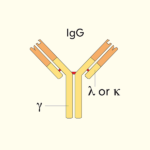
体内实验级重组抗生物素(Anti-biotin)单克隆抗体,克隆号 F1,恒定区为小鼠 IgG2a Kappa(Mouse IgG2a Kappa),由哺乳动物细胞重组表达。该抗体识别共价偶联在蛋白质、多肽、核酸或固相基质上的生物素分子,可作为第二步检测试剂用于各类生物素化靶标的检测。生物素-链霉亲和素系统因亲和力极高被广泛用于免疫检测信号放大;但由于生物素天然存在于多种生物样本,会造成灵敏度下降、假阴性或假阳性,抗生物素抗体与链霉亲和素/生物素封闭试剂可有效降低内源性生物素及生物素结合蛋白带来的背景干扰。本品经 Protein G 亲和纯化,还原性 SDS-PAGE 纯度>95%,LAL 法内毒素<1 EU/mg,0.2 μM 过滤于 1× PBS,适用于 ELISA、WB、流式细胞术、免疫沉淀、石蜡/冰冻切片 IHC 及体内动物模型研究;小鼠 IgG2a 同种型对小鼠 Fcγ 受体(含激活型 FcγRIV)结合能力较强,适合需要效应功能的体内实验,亦可提供荧光素、酶标记等偶联版本及 Avi-/His-/DYKDDDDK 标签突变体。
产品参数
| 货号 | PA007522.m2a |
|---|---|
| 产品名称 | 重组抗生物素抗体,小鼠IgG2a Kappa | Syd Labs PA007522.m2a |
| 英文名 | In vivo Grade Recombinant Anti-biotin Antibody, Mouse IgG2a Kappa |
| 供货商名称 | Syd Labs, Inc. |
| 品牌名 | Syd Labs |
| 别称 | 生物素,维生素H,维生素B7 |
| 克隆号 | F1 |
| 同种型 | 小鼠IgG2a Kappa |
| 特异性 | 该体内实验级重组小鼠单克隆抗体(克隆F1)特异性结合生物素(Biotin,维生素H/维生素B7),可识别偶联于蛋白质、多肽、核酸或固相基质上的生物素 |
| 应用 | ELISA、Western Blot(WB)、流式细胞术(Flow)、免疫沉淀(IP)、免疫组化-石蜡切片(IHC-P)、免疫组化-冰冻切片(IHC-F)及体内动物模型研究 |
| 抗体形式 | 0.2 μM过滤溶液,1x PBS |
| 内毒素 | 根据 LAL 方法,≤ 1 EU每 1 mg 蛋白质 |
| 纯度 | >95%(在还原条件下通过SDS-PAGE测定) |
| 运输 | 体内实验级重组抗生物素抗体(克隆F1)采用冰袋运输;收到后请立即按推荐温度保存 |
| 稳定性与存储 | 使用手动除霜冰箱并避免重复冻融循环。 自收到之日起 1个月,保存在2 至 8°C。 自收到之日起 12个月,保存在-20°至-70°C。 |
| 产品咨询 | Syd Labs在国内只通过代理商销售其产品,不做直销。终端用户咨询价格请联系Syd Labs中国代理商。 关于Syd Labs产品如果有任何技术或其它问题,欢迎随时联系Syd Labs国内市场推广合作伙伴:武汉多找找科技有限公司,企业微信:duozhaozhao2024 联系电话:18162581039(龙经理) |
| 应用详情 | ELISA、Western Blot(WB)、流式细胞术(Flow)、免疫沉淀(IP)、免疫组化-石蜡切片(IHC-P)、免疫组化-冰冻切片(IHC-F)及体内动物模型研究 |
文献
PA007522.m2a:体内实验级重组抗生物素单克隆抗体,小鼠IgG2a Kappa(In vivo Grade Recombinant Anti-biotin Antibody, Mouse IgG2a Kappa)
重组抗生物素抗体,小鼠IgG2a Kappa背景知识
生物素抗体(重组小鼠抗生物素抗体) 可特异性识别结合在蛋白质、多肽、核酸或固相基质上的生物素(维生素B7),并提供荧光基团/酶偶联版本的生物素抗体。
该抗生物素单克隆抗体可作为二级检测试剂,用于多种生物素化靶标的分析应用。由于链霉亲和素与生物素具有极高的亲和力,生物素-链霉亲和素系统被广泛应用于免疫检测信号放大技术。
鉴于生物素天然存在于多种生物样本中,其可能影响基于生物素的检测结果(如灵敏度降低、假阴性或假阳性)。当采用基于生物素的免疫组化(IHC)检测时,需使用链霉亲和素/生物素封闭试剂盒、亲和素/生物素封闭系统或生物素封闭剂,以阻断组织内源性亲和素、生物素及生物素结合蛋白的信号干扰。
重组抗生物素抗体,小鼠IgG2a Kappa部分参考文献:
1.Determination of the epitope specificities of monoclonal antibodies using unprocessed supernatants of hybridoma cultures
Würzner R, Oppermann M, Zierz R, Baumgarten H, Götze O
J Immunol Methods. 1990;129(2):245–252. PMID: 1689363 DOI: 10.1016/0022-1759(90)90155-O
“A double monoclonal antibody (mAb) ELISA has been developed to determine the epitope specificities of murine monoclonal antibodies (mAbs). It permits the mAbs which bind to the same or adjacent epitopes to be distinguished from those which bind to separate epitopes on soluble monomeric proteins. The assay is designed for the early evaluation of mAbs in hybridoma culture supernatants when purified or labeled mAbs are not available. It does not rely on chemical or enzymatic fragmentation of the antigen and does not generate results which may be due to differences in the affinities of the mAbs. The test was evaluated using culture supernatants from hybridomas which produced mAbs against the complement proteins C3a, C6 or C7, with biotin-conjugated antibodies as key detection reagents, providing a robust framework for biotin-based mAb characterization.”
2.The effect of biotinylation on the antigenic specificity of anti-defensin monoclonal antibodies
Panyutich AV, Baturevich EA, Kolesnikova TS, Ganz T
J Immunol Methods. 1993;164(2):195–200. PMID: 7679133 DOI: 10.1016/0022-1759(93)90219-W
“We studied the effects of biotinylation on three monoclonal antibodies (Mabs) that were raised against carrier protein conjugates of human defensin HNP-1, and of rabbit defensins NP-2 and NP-5 respectively. Before biotinylation, each Mab specifically bound to its peptide hapten. Biotinylation of these Mabs by the N-hydroxysuccinimide-biotin (NHS-biotin) resulted in crossreactivity of each Mab with the two irrelevant defensin peptides. In contrast, Mab specificity was preserved after biotinylation with biotin hydrazide, which links biotin to the glycan moiety of antibodies. These results highlight important considerations for biotin-based detection systems and the proper selection of anti-biotin or streptavidin reagents in immunoassays.”
3.Biotin interference in clinical immunoassays: an update
Li D, Ferguson A, Cervinski MA, Lynch KL, Kyle PB
Clin Biochem. 2017;50(18):1155–1163. PMID: 28355359 DOI: 10.1016/j.clinbiochem.2017.03.017
“Biotin is an essential water-soluble B vitamin that has gained popularity in high-dose oral supplementation for cosmetic and medical purposes, including treatment of multiple sclerosis. High-dose biotin supplementation can cause clinically significant interference with biotin-streptavidin-based immunoassays, leading to falsely low or high results depending on assay design. This review summarizes the mechanisms of biotin interference, affected assays (troponin, TSH, thyroid hormones, tumor markers, etc.), and mitigation strategies, including the use of anti-biotin antibodies and biotin-blocking reagents to neutralize free biotin in specimens.”
4.Biotin and streptavidin in diagnostics and biotechnology: past, present and future
Dundas CM, Demonte D, Park S
Appl Microbiol Biotechnol. 2013;97(21):9343–9353. PMCID: PMC2440739(注:PMCID请以实际收录为准) PMID: 23892738 DOI: 10.1007/s00253-013-5232-7
“The high-affinity interaction between biotin and streptavidin/avidin has been exploited for decades in diagnostics, affinity purification, imaging and targeted delivery. This review covers the molecular basis of the biotin-streptavidin interaction, common biotinylation chemistries, assay formats (ELISA, WB, IHC, flow cytometry), and the challenge of endogenous biotin background in tissues. We discuss the use of monoclonal anti-biotin antibodies as alternative detection reagents to streptavidin when lower background or biotin-competition resistance is required in IHC and other immunoassays.”
5.The use of endogenous biotin-blocking reagents in immunohistochemistry: a comparative study
Varga VS, Bocsi J, Sipos F, Tulassay Z, Molnar B
Appl Immunohistochem Mol Morphol. 2003;11(1):74–79. PMID: 12659070 DOI: 10.1097/00129039-200303000-00011
“Endogenous biotin is a well-known source of false-positive staining in immunohistochemistry, particularly in mitochondria-rich tissues such as kidney, liver, heart and steroid-producing cells. This study compares the effectiveness of avidin/biotin blocking kits versus monoclonal anti-biotin antibody-based blocking strategies in eliminating streptavidin–biotin background in formalin-fixed paraffin-embedded tissues. The results show that anti-biotin antibodies can provide equivalent or superior blocking of endogenous biotin epitopes with less non-specific binding compared to avidin-based blockers, supporting their use as an alternative blocking reagent in diagnostic and research IHC.”


